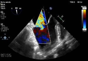
YOU BE THE CARDIAC IMAGING SPECIALIST!
What abnormality is demonstrated in this color flow Doppler echocardiogram?
ANSWER
Functional Mitral Regurgitation!
In the presence of heart disease, the mitral annulus can dilate to the point where the leaflets of the mitral valve no longer meet to create a seal (coapt). When this happens, blood can flow backward into the atrium during cardiac systole, creating mitral regurgitation. In the absence of frank pathology to the mitral valve itself, this form of backward flow is called Functional Mitral Regurgitation (FMR).
This FMR was seen in a sheep and this image of the heart was acquired using IMMR’s X8-2t TEE probe on an EPIQ machine, with great spatial and temporal resolution.
Mitral regurgitation (MR) can result from various abnormalities of the mitral valve. It is the most common valvular abnormality worldwide, affecting over 2% of the total population. Severe mitral valve regurgitation can lead to complications, including heart failure, atrial fibrillation and pulmonary hypertension. Treatment of MR with mitral valve repair or replacement devices is often indicated.
Large animal models are invaluable in MedTech research for the investigation of the pathophysiology of mitral regurgitation. When it comes to choose the relevant animal models for preclinical validation of novel mitral devices, intimate knowledge of comparative anatomy is a key competency for obtaining meaningful results.
Contact us to learn more and discuss your preclinical research and pathology needs.

