You be the Imaging Specialist
Which state-of-the-art technology provides detailed images like this one and enables you to tailor your vascular studies, analyze your results with high precision and advance your product development?
ANSWER
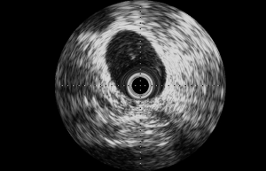
This cross-sectional view of a stent in the external iliac vein of a sheep that was acquired at Veranex is a beautiful example of images provided by intravascular ultrasound (IVUS)!
IVUS is a medical imaging modality using a miniaturized ultrasound probe attached to the distal end of a specialized catheter that provides a 360° view from inside blood vessels out through the surrounding blood column and to visualize the endothelium. This technique has contributed to technological improvements in the diagnosis and treatment of coronary, peripheral and cerebral vascular disorders.
When conducting vascular preclinical research, IVUS may be a valuable tool for evaluation of vessel morphology and size before implantation, guidance of device implantation and assessment of long-term device position and safety.
A complementary technique, Optical Coherence Tomography (OCT), is an imaging modality that uses near-infrared light to provide high-definition images with higher spatial resolution and tissue penetration than IVUS. Both OCT and IVUS are available at Veranex, and our imaging specialists can help you determine which technology is best for your needs.
If you are interested in learning more about our preclinical research and pathology services, we encourage you to get in touch with us. We would be delighted to discuss your specific needs and plans.