You be the Cardiac Imaging Specialist!
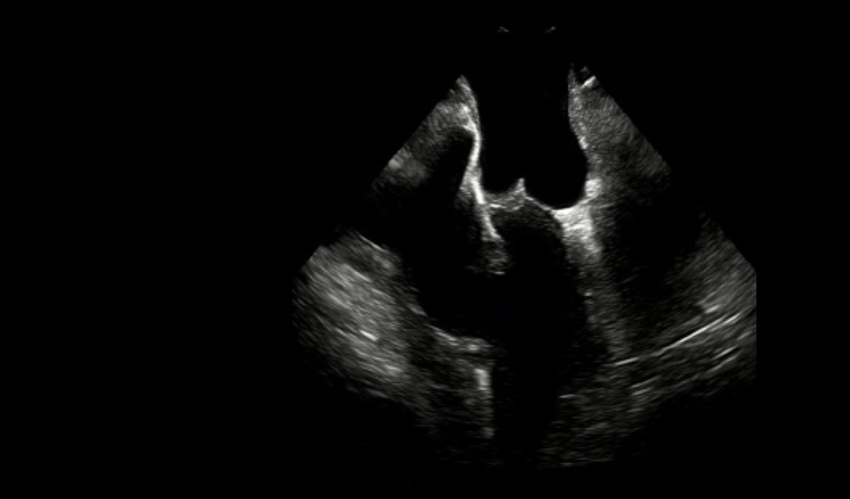
What cardiac structures are observable on this IntraCardiac Echocardiography (ICE) image, and what approach was used to demonstrate them?
Answer:
The left atrium, left ventricle and aorta are the cardiac structures shown, and this ICE image was obtained with an arterial approach, with the ICE catheter positioned in the aortic arch!
Intracardiac ultrasound is designed for real-time image guidance and visualization of cardiac anatomy. It can be instrumental in a variety of diagnostic and therapeutic interventions in structural heart disease, electrophysiology and other cardiac interventions.
Veranex remains at the forefront of innovative imaging, and we routinely utilize diverse advanced echocardiographic techniques for intraprocedural cardiovascular visualization. These include Transthoracic (TTE), Trans-Esophageal (TEE), Epicardial, Transdiaphragmatic, and Intracardiac (ICE) Echocardiography.
This high-definition ICE image of cardiac anatomy was acquired using Veranex’s state-of-the-art St Jude ViewFlex™ ICE 9-French catheter and Zonare ViewMate™ Z Intracardiac Ultrasound console.
If you are interested in learning more about our preclinical research and pathology services, we encourage you to get in touch with us. We would be delighted to discuss your specific needs and plans.
