You be the Pathologist!
What type of implant is shown here, and in what location?
Answer:
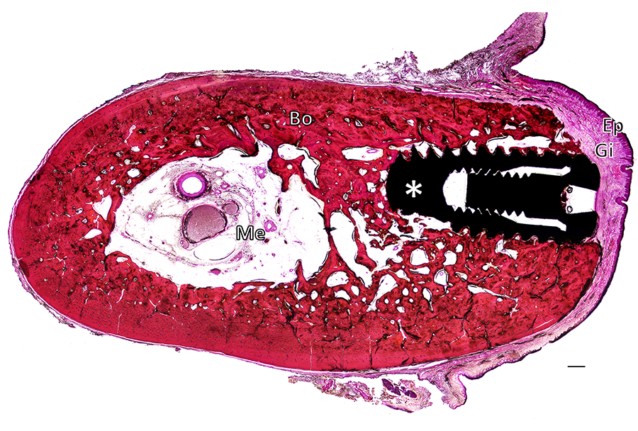
This is a dental implant in the mandible!
This mandibular dental implant was evaluated microscopically 34 days after implantation. Using polymethyl methacrylate (PMMA) resin embedding, our Board-certified Veterinary Pathologists can assess the healing and integration of the implant in situ. As shown here, the gingiva (Gi) and its epithelium (Ep) are perfectly sealed, and the implant (white asterisk) is completely integrated in mature bone tissue (Bo).
H&E, original magnification: x1, scale bar: 1 mm.
Ep: Epithelium. Gi: Gingiva. Bo: Mandibular bone. Me: Medullary cavity. White asterisk: Dental implant.
This histopathology image is one example from the comprehensive suite of Pathology Services offered by Veranex Preclinical Services’ in-house team of Veterinary Pathologists.
If you are interested in learning more about our preclinical research and pathology services, we encourage you to get in touch with us. We would be delighted to discuss your specific needs and plans.
